Blog
Window to the Nano World



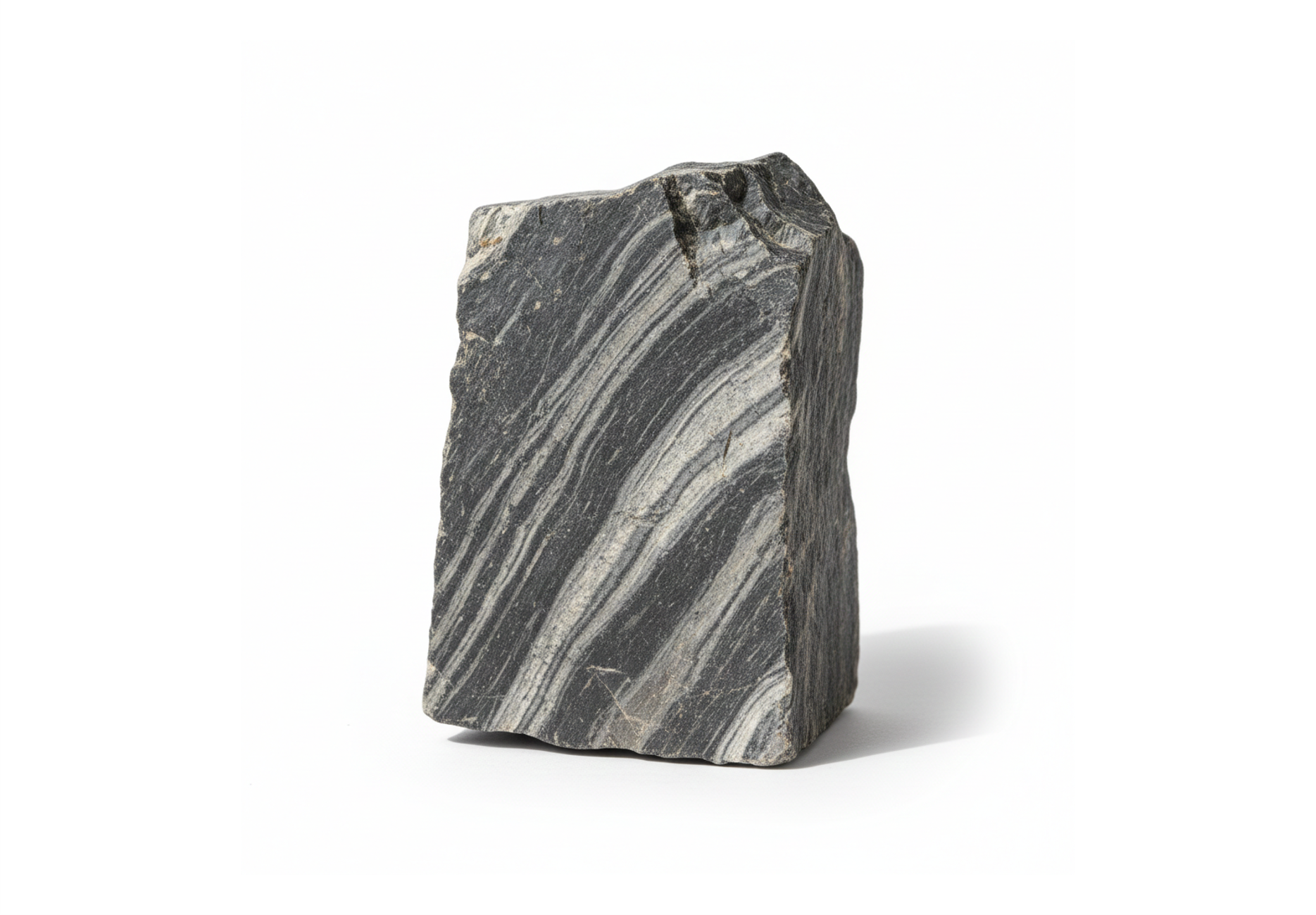
The rock industry has played a fundamental role in human civilization, supplying natural stone materials for construction, manufacturing, and industrial applications. Common rock types such as granite, marble, limestone, and basalt are extracted through quarrying and processed through cutting, crushing, and polishing for various end uses.
Processed rocks are widely used as construction aggregates, architectural materials, and essential raw materials in industries such as cement, glass, and steel production. To accurately evaluate rock quality, detailed analysis of mineral particle size, morphology, distribution, and physical characteristics is essential — making electron microscopy a powerful analytical tool in geological and material studies.
COXEM’s CX Series SEM is well suited for rock and mineral analysis thanks to its large chamber design, which accommodates samples of various sizes and enables fast, convenient observation of multiple specimens. Its rapid vacuum system and multiple vacuum modes also allow stable imaging of porous samples without difficulty.
In addition, the CX Series can be integrated with analytical tools such as EDS and EBSD, enabling qualitative and quantitative elemental analysis of minerals as well as crystallographic analysis for evaluating material strength and fracture behavior.
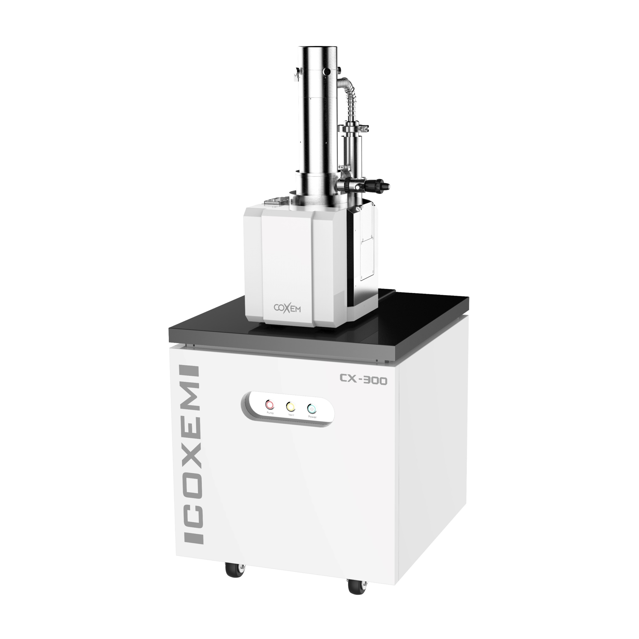
Samples compatible with the CX Series should be smaller than 100 mm × 100 mm × 60 mm. If the rock specimen is too large, it should be cut or mechanically polished to fit within the chamber size limitations.
Since most natural rocks are non-conductive, conductive coating is typically required for stable SEM observation. For elemental analysis, it is recommended to use a coating material that is not present in the sample composition to avoid interference with qualitative or quantitative results.
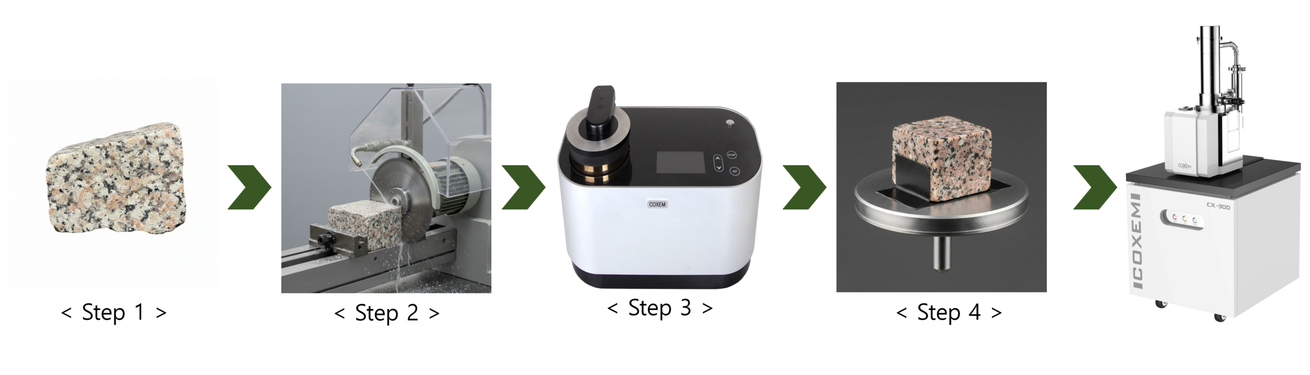
Step 1. Prepare the rock sample for observation.
Step 2. If the sample is too large, reduce its size by cutting or mechanical polishing.
Step 3. Place the sample into the SPT-20 coater and apply a Pt or Au conductive coating.
Step 4. Mount the sample onto the SEM holder using carbon tape or silver paste.
Step 5. Load the sample into the CX-300 and perform SEM observation.

[Rock(SE)]

[Rock(BSE)]
The rock sample was observed at 15 kV using both SE and BSE imaging modes. In SE mode, the surface morphology of the rock could be visualized in detail, allowing cracks and surface defects to be clearly identified.
When the same area was observed in BSE mode, differences in image contrast appeared according to the elemental composition of the minerals, enabling clear visualization of compositional distribution within the rock sample.
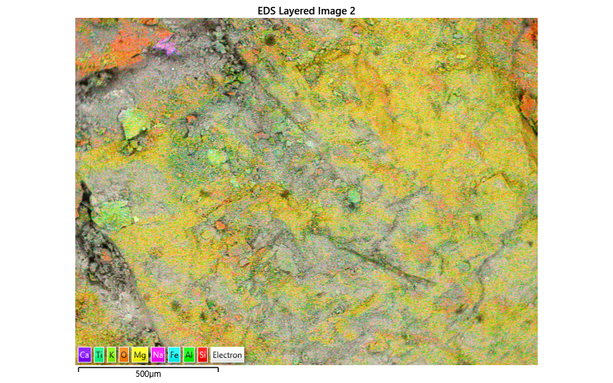
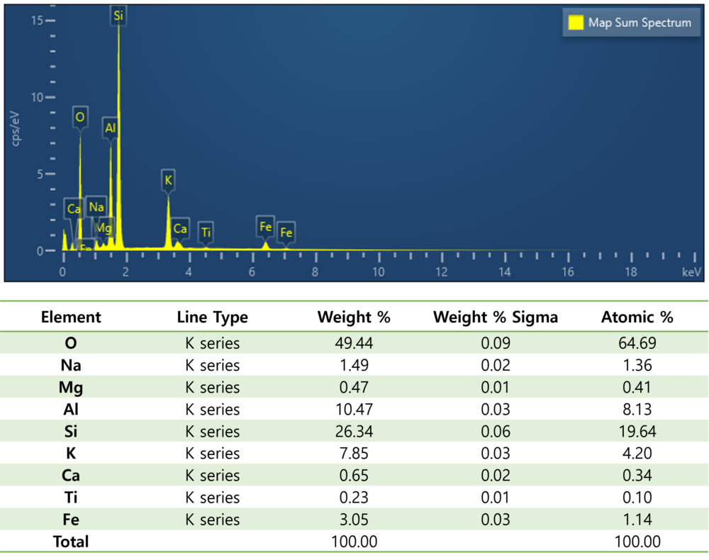
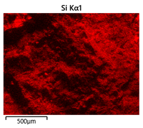
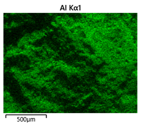

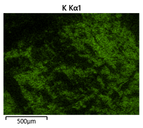
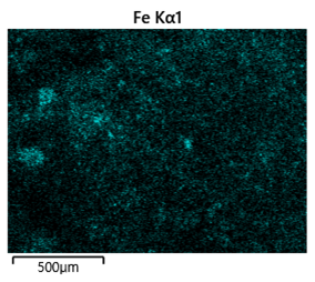

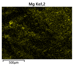
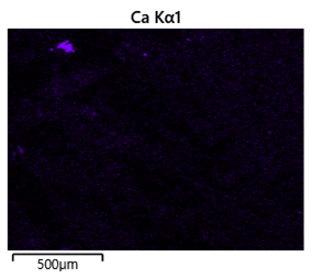
Elemental mapping analysis was performed on the rock surface to examine the distribution of detected elements. Silicon, aluminum, and oxygen were predominantly detected across the surface, while smaller amounts of elements such as potassium, sodium, and iron were also identified.
# Rock #Materials Sceience #SEM #CX-300 #Ionsputtercoater #SPT-20

 Discovering the Hidden World of Insects with SEM