Blog
Window to the Nano World




Scanning Electron Microscopy (SEM) is widely used in life science research to investigate the structure, composition, and ecological characteristics of living organisms at the micro- and nanoscale. Because SEM uses an electron beam with a much shorter wavelength than visible light, it can reveal ultra-fine structures that cannot be observed with the naked eye or conventional optical microscopes.
This capability allows researchers to study a wide range of biological samples, from microorganisms and fungi to insects, plant tissues, and nanoscale algae, with exceptional detail.
SEM has become an essential analytical tool for observing the microscopic world of life. Surface structures such as fungal hyphae, bacterial cell walls, insect compound eyes, and plant stomata can be visualized at high resolution, supporting species identification and growth-stage analysis.
SEM is also highly valuable for studying structural damage and biological responses caused by antimicrobial agents, contaminants, or environmental changes, making it useful in food safety, environmental science, and pathogen research. When combined with EDS, SEM can further analyze elemental composition, such as silicon in diatoms or metallic elements on insect surfaces.
COXEM’s Tabletop SEM systems provide an additional advantage through Low Vacuum (LV) imaging, enabling observation of uncoated biological samples while preserving their natural state.
Proper SEM sample preparation for biological specimens focuses on preserving delicate microstructures with minimal deformation. Samples are typically fixed using 2.5% glutaraldehyde to stabilize cellular structures, followed by gradual ethanol dehydration (30 → 100%) to remove moisture while minimizing structural shrinkage.
The dehydrated sample is then fully dried using HMDS substitution or freeze-drying methods. After mounting onto an SEM stub, Pt or Au sputter coating is applied to improve conductivity and reduce charging effects during SEM observation.
This preparation workflow enables high-resolution SEM imaging while maintaining the original morphology of biological samples as much as possible.
Step 1. Store the biological sample at room temperature while minimizing physical damage.
Step 2. Treat the sample sequentially using step-by-step preparation solutions.
Step 3. Mount the stabilized sample onto the SEM stub.
Step 4. Apply Au sputter coating using the SPT-20 to ensure stable SEM observation.


[Zebrafish]

[Mosquito]
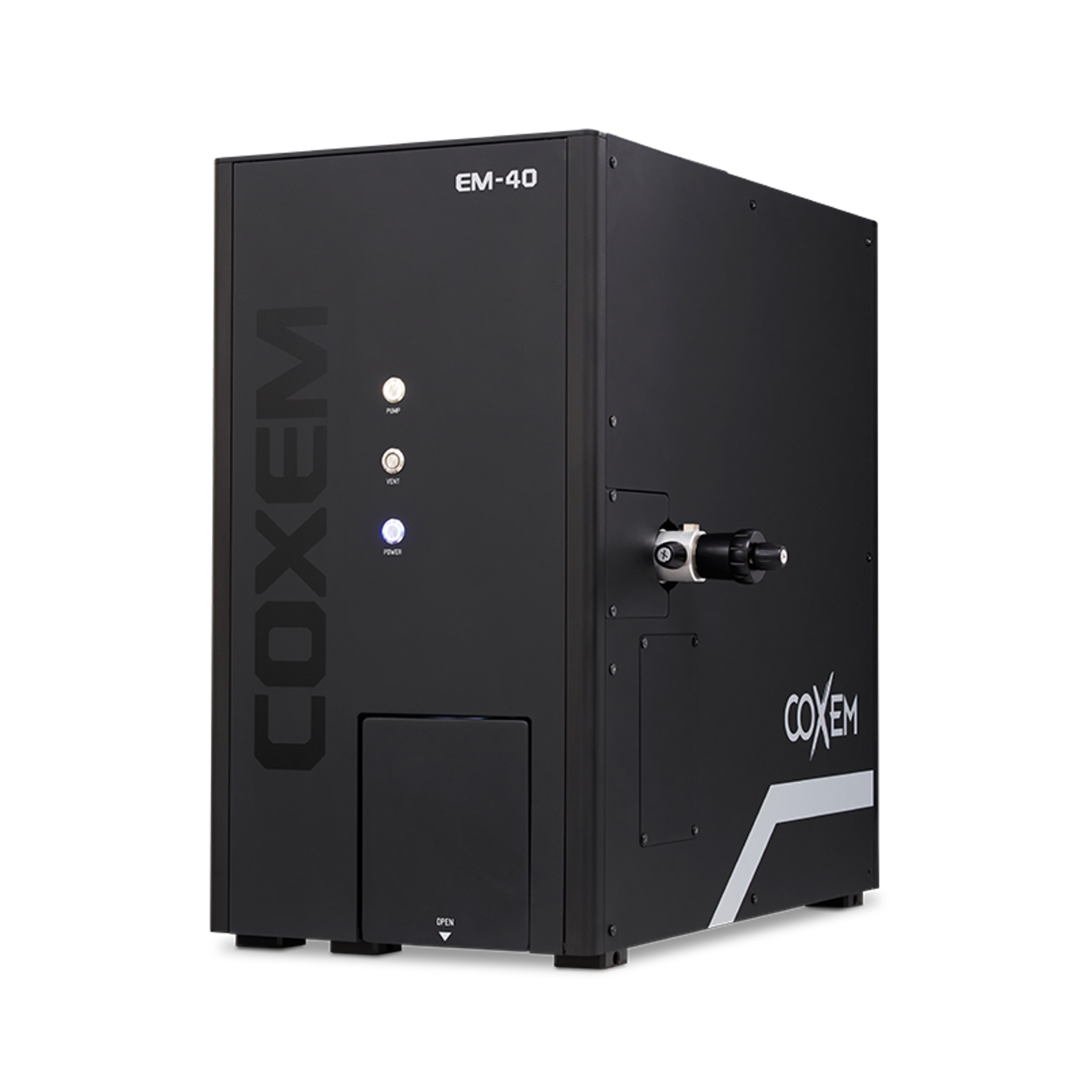
The analysis was performed using the COXEM EM-40 at an accelerating voltage of 15 kV and a working distance of 12 mm using both SE and BSE detectors.
By performing proper biological sample preparation prior to SEM observation, clearer and more stable SEM images can be obtained with improved surface detail and structural visibility.

[Bee’s Head]

[Bee’s Head]
These images were acquired using the COXEM EM-40 scanning electron microscope. Structures on the bee’s head that appeared fiber-like at low magnification revealed well-defined branch-like morphologies when observed at higher magnification.

[Larva]

[Caterpillar]

[Chrysanthemum flower]

 The Hidden Microstructure of Everyday Straws