Blog
Window to the Nano World




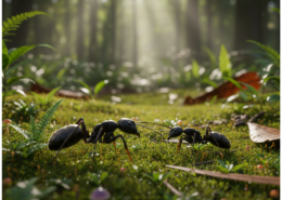
Insects make up the majority of Earth’s species and play a vital role in ecosystems and material circulation. Although they may appear simple externally, their structures — including exoskeletons, wings, and sensory organs — are highly optimized for efficiency and functionality.
For example, insect exoskeletons feature lightweight yet strong layered structures that inspire the development of advanced materials in the aerospace and automotive industries. The microscopic hair structures on a fly’s foot provide remarkable adhesion without glue, influencing technologies such as medical patches and bio-inspired robotics. These unique biological features continue to inspire innovation across a wide range of industries.
However, the nanoscale and microscale structures found on insect surfaces cannot be fully analyzed using optical microscopes alone. SEM is essential for revealing these fine structural details with high resolution.
For fast and convenient biological sample observation, COXEM recommends the EM-40 tabletop SEM. In addition to SE and BSE imaging modes, optional features such as Low Vacuum mode and a Cooling Stage enable stable observation of delicate biological samples while minimizing sample damage.
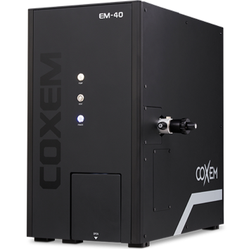
When observing biological samples with SEM, moisture must be completely removed to ensure stable imaging under vacuum conditions. Samples should therefore be thoroughly dried using a hot plate or drying oven before observation. Since biological specimens are generally non-conductive, a thin conductive coating such as platinum or gold is typically applied to reduce charging during SEM imaging.
If conductive coating is not preferred, Low Vacuum (LV) mode with BSE imaging can be used to minimize charging effects while preserving the sample surface.
Step 1. Collect the sample.
Step 2. Thoroughly dry the sample to remove residual moisture.
Step 3. Place the sample into the SPT-20 coater, select the target material, and coat at 4 mA for 300 seconds.
Step 4. Mount the coated sample onto the sample holder.
Step 5. Load the sample into the EM-40 and perform SEM observation.

[Bee Eye]

[Bee Head]
When observing biological samples with SEM, reducing both the accelerating voltage and electron beam size is recommended to minimize sample damage. High-magnification imaging of a bee’s face revealed uniformly arranged hexagonal compound eyes measuring several tens of micrometers, along with densely distributed cylindrical hairs on the facial surface.
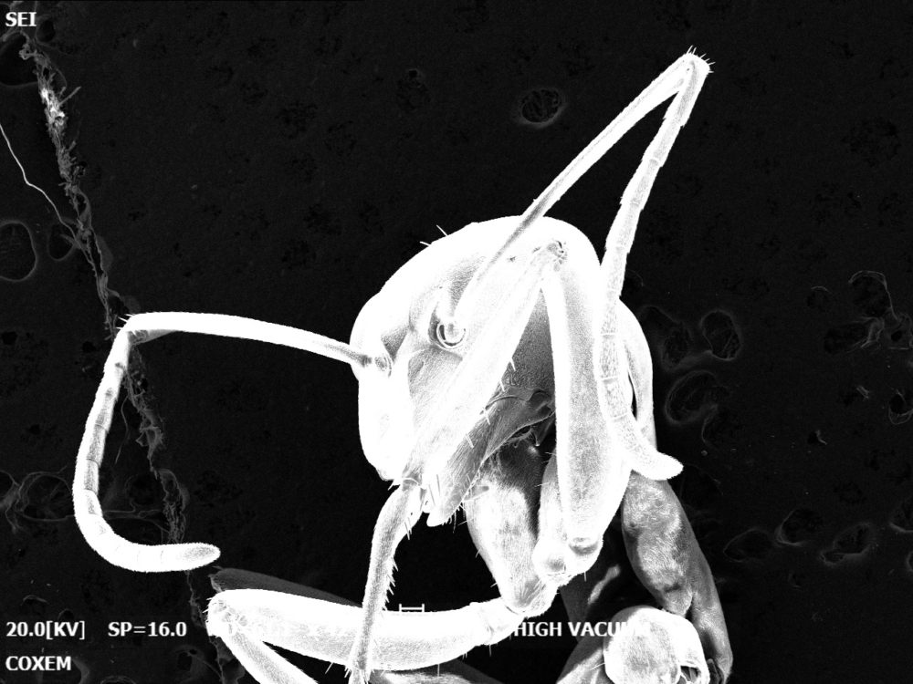
[Ant Head, High Vacuum]
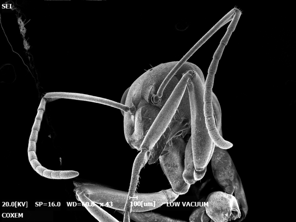
[Ant Head, High Vacuum]
When non-conductive samples are observed without conductive coating, electrons can accumulate on the surface, causing a charging effect. Severe charging may lead to abnormal brightness fluctuations during imaging or even damage to the sample, as shown in the image above.
Using Low Vacuum (LV) mode helps reduce charging by introducing gas molecules into the chamber, which neutralize accumulated surface charges. This enables stable observation of uncoated non-conductive samples with improved imaging reliability.
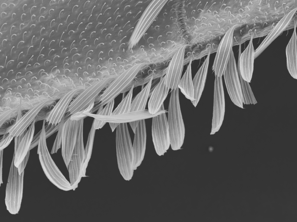
[Fly Wing]
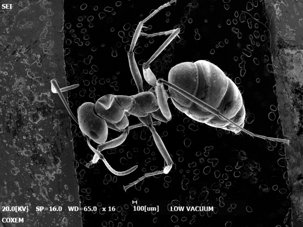
[Ant]
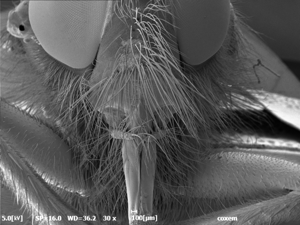
[Bee Head]
#Insects #LifeScience #Bio #EM-40 #LowVacuum

 Precise Cross-Section Analysis of MLCC Structures