Blog
Window to the Nano World


Scanning Electron Microscopy (SEM) is an essential tool in dental material research, offering detailed visualization of surface microstructures that influence biological interactions. In this article, we report a comparative surface analysis of natural permanent tooth roots and commercial dental implants using the COXEM EM-40 Tabletop SEM. Results highlight the naturally irregular and porous morphology of tooth roots compared with the engineered, uniformly roughened surfaces of implants. These findings emphasize the importance of SEM in understanding functional differences and provide insights into how COXEM’s EM-40 can serve as a practical solution for both academic research and clinical studies.
Keywords: SEM, EM-40, tooth root, dental implant, surface analysis, osseointegration
Dental health plays a vital role not only in daily functions such as mastication but also in overall well-being and quality of life. Natural permanent teeth and dental implants may serve similar purposes, but their surface characteristics reveal important differences that directly affect their biological functions.
The permanent tooth root connects with surrounding periodontal ligaments, allowing the tooth to withstand mechanical forces while providing shock absorption. By contrast, dental implants must integrate directly with bone tissue through a process known as osseointegration. This integration is highly dependent on the surface microstructure of the implant, which is intentionally engineered to optimize bone adhesion.
Visualizing these microstructural differences requires advanced imaging technology. Scanning Electron Microscopy (SEM) provides high-resolution surface imaging, making it indispensable in dental research. In this study, we employed the COXEM EM-40 Tabletop SEM, a compact and user-friendly instrument that combines accessibility with robust imaging performance, to compare the surfaces of permanent tooth roots and dental implants.
2.1 Instrumentation
The analysis was carried out using the COXEM EM-40 Tabletop SEM, a compact yet versatile system capable of generating detailed surface images at magnifications up to 50,000×. Its intuitive interface and streamlined sample preparation requirements make it suitable for both educational and advanced research environments.
2.2 Experimental Conditions
2.3 Samples Preparation
3.1 Permanent Tooth Root Surface
SEM imaging revealed that the tooth root surface exhibits a highly irregular and porous morphology. Micro-pores of varying sizes and shapes were visible, along with complex textures that suggest natural wear and mineral deposition over time.
From a functional perspective, these surface irregularities provide anchoring sites for periodontal ligament fibers, which play a critical role in stabilizing the tooth within the alveolar bone. Moreover, the porous structure contributes to shock absorption during chewing, allowing the tooth to withstand significant mechanical forces.

Fig (a)
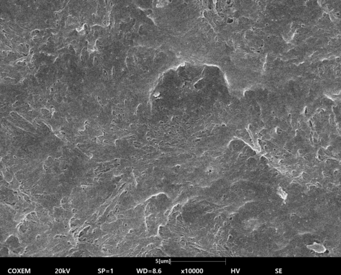
Fig (c)

Fig (b)
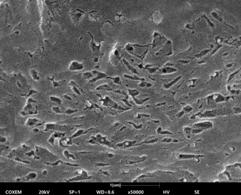
Fig (d)
Fig (a)-(d). SEM Images of Permanent Tooth Root Surface
3.2 Dental Implant Surface
The implant surface displayed a controlled, uniformly roughened microstructure created by sandblasting and acid-etching treatments. Under SEM, the surface appeared covered with evenly distributed pits and ridges, offering a reproducible and predictable topography.
These features are specifically engineered to promote cell adhesion and enhance osseointegration. The uniformity of the roughness ensures that bone cells can attach consistently across the implant surface, improving the likelihood of long-term stability and reducing the risk of implant failure.
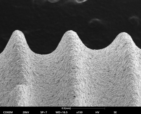
Fig (e)

Fig (g)
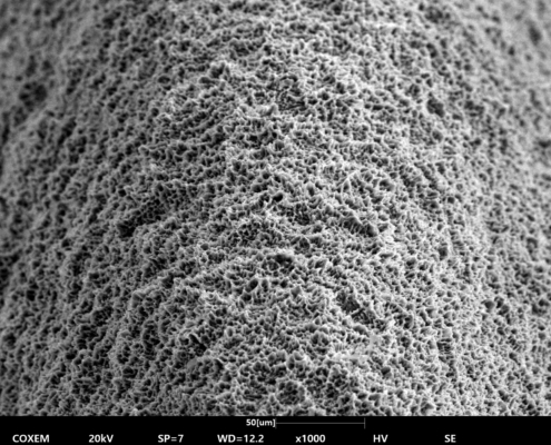
Fig (f)

Fig (h)
Fig (e)-(h). SEM Images of Dental Implant Surface
3.3 Discussion
The comparative SEM analysis demonstrates that natural teeth and dental implants employ fundamentally different mechanisms for stability.
This analysis highlights the value of SEM imaging in dental material research. By directly visualizing surface features, SEM provides insights into how microstructural design translates into biological performance.
The COXEM EM-40 Tabletop SEM is particularly well-suited for such investigations. Its compact design allows installation even in small laboratories, while its performance rivals larger, more complex SEM systems. Furthermore, with the optional integration of EDS (Energy Dispersive Spectroscopy), compositional analysis of dental materials becomes possible, adding another layer of insight.
The surface comparison between permanent tooth roots and dental implants underscores the complementary strategies of natural and artificial structures:
Through this study, the COXEM EM-40 Tabletop SEM has proven its capability as a powerful yet accessible tool for visualizing such differences. Its combination of simplicity, high-resolution imaging, and analytical versatility makes it an excellent choice for dental researchers, material scientists, and clinicians seeking to understand and optimize dental solutions.
COXEM will continue to support advancements in biomaterial research by providing reliable SEM solutions that bridge the gap between natural biology and engineered technology.

 Rice Cake