Blog
Window to the Nano World


Cherry blossoms are widely recognized for their delicate beauty and symbolic representation of spring. While their macroscopic appearance appears soft and uniform to the naked eye, their microstructure reveals a far more complex and functional architecture.
Petals that appear smooth, pollen perceived as fine particles, and seemingly simple leaf surfaces are all composed of intricate microstructures designed to perform specific biological functions.
In this study, COXEM’s Tabletop Scanning Electron Microscope (EM-40) was utilized to observe and analyze the microstructures of cherry blossom petals, pollen, and leaf surfaces. This approach enables a deeper understanding of the functional characteristics of natural materials through high-resolution imaging.

Samples including cherry blossom leaves, petals, and pollen were observed using the COXEM EM-40 Tabletop SEM.

The SEM analysis enabled detailed visualization of microstructures that are not observable through conventional optical methods, including:
SEM analysis was performed using the COXEM EM-40 Tabletop Scanning Electron Microscope under the following conditions:
These conditions enabled clear visualization of compositional contrast and internal structural features.
SEM observations revealed distinct microstructural characteristics across different parts of the cherry blossom.
Leaf Surface (Fig. a, b)
The leaf surface consists of stomata, micro-protrusions, and a wax layer. These structures serve multiple functional roles including:
Additionally, these features contribute to environmental adaptability and enable self-cleaning functionality.
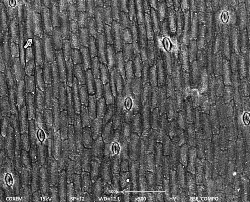
Fig. (a)
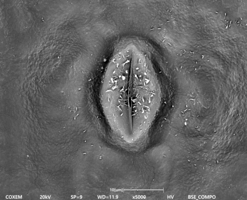
Fig. (b)
Petal Surface (Fig. c, d)
Unlike its smooth visual appearance, the petal surface is composed of fine cellular structures.
These microstructures scatter light, contributing to:
This represents a typical example of structural coloration in natural materials.

Fig. (c)
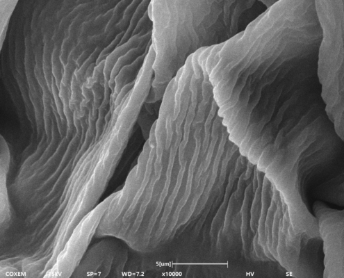
Fig. (d)
Pollen Structure (Fig. e, f)
Pollen particles exhibit near-spherical shapes with textured surfaces including protrusions and network-like patterns.
These morphological features:
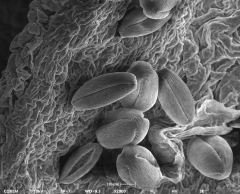
Fig. (e)
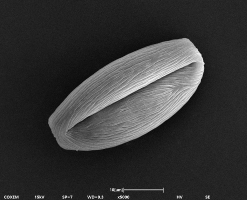
Fig. (f)
COXEM provides advanced microstructural analysis solutions based on Tabletop SEM technology.
Through high-resolution imaging, users can:
This enables efficient and insightful analysis across a wide range of applications, from biological samples to industrial materials.
This study demonstrates that Tabletop SEM analysis provides a powerful method for understanding the microstructural and functional characteristics of natural materials such as cherry blossoms.
By utilizing the COXEM EM-40:
Ultimately, COXEM’s SEM solution offers an effective platform for uncovering hidden structures and enabling intuitive, high-quality microstructural analysis.
#SEM #Tabletopsem #EM-40 #Lifescience #BSE

 Cross Section of Solder Ball