Blog
Window to the Nano World



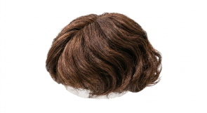
Human hair is a protein-based fiber composed of several distinct structural layers. The cuticle forms the outermost protective layer, shielding the hair from physical damage and chemical exposure while also influencing texture and shine. It consists of thin, overlapping keratin cells arranged like scales.
Keratin contains cysteine, a sulfur-containing amino acid that forms strong disulfide bonds, giving hair its mechanical strength and durability. Hair treatments such as dyeing and perming work by breaking and reforming these disulfide bonds. Beneath the cuticle lies the cortex, which makes up most of the hair thickness, while the medulla forms the central core and may contain air spaces and cellular residues that influence hair thickness and permeability.
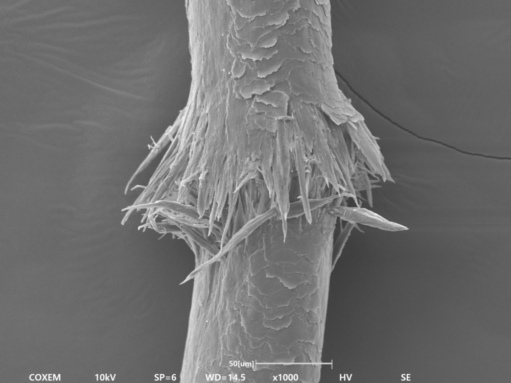
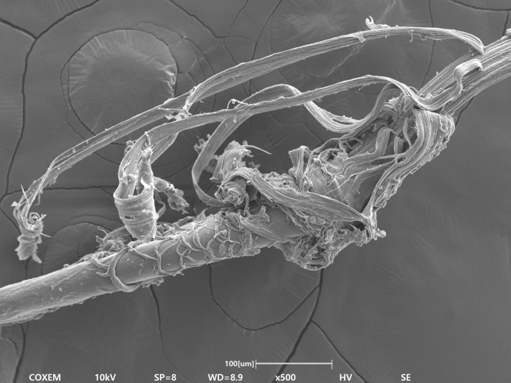
To analyze these fine structures in detail, SEM observation is essential. Features such as cuticle overlap, surface cracks, and pore structures can be examined at high resolution, making SEM highly valuable for hair damage assessment and cosmetic product development.
COXEM’s EM-40 tabletop SEM enables high-resolution imaging down to the nanometer scale, allowing detailed observation of cuticle morphology and damaged regions on the hair surface. Equipped with both SE and BSE detectors and compatible with compact EDS integration, the EM-40 supports not only surface morphology observation but also elemental distribution and compositional analysis.

Human hair is a non-conductive material, so direct SEM observation without conductive coating can cause severe charging effects, making stable imaging difficult. Hair is also sensitive to heat, meaning prolonged exposure to the electron beam may damage the sample surface.
To minimize charging and prevent beam-induced damage, conductive coating is strongly recommended before SEM analysis. If coating is not preferred, observation under Variable Pressure (VP) mode is recommended for more stable imaging.
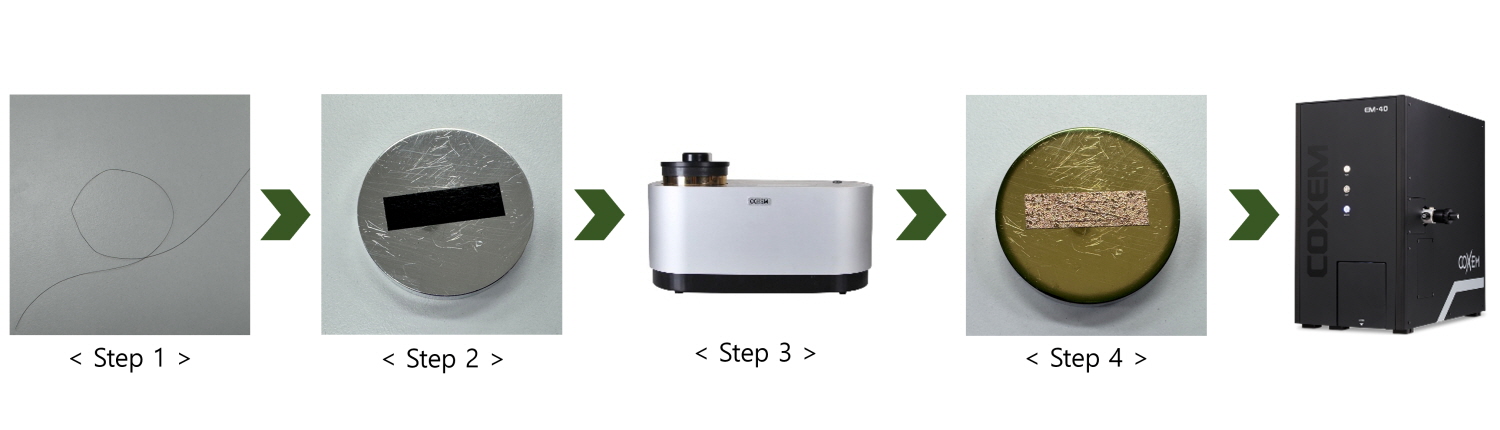
Step 1. Collect and cut the hair sample to an appropriate size.
Step 2. Mount the hair onto the sample holder using carbon tape.
Step 3. Place the mounted sample into the SPT-20 sputter coater and apply Pt or Au coating.
Step 4. After coating, load the sample into the EM-40, evacuate the chamber, and observe the hair surface.

[Hair 1]
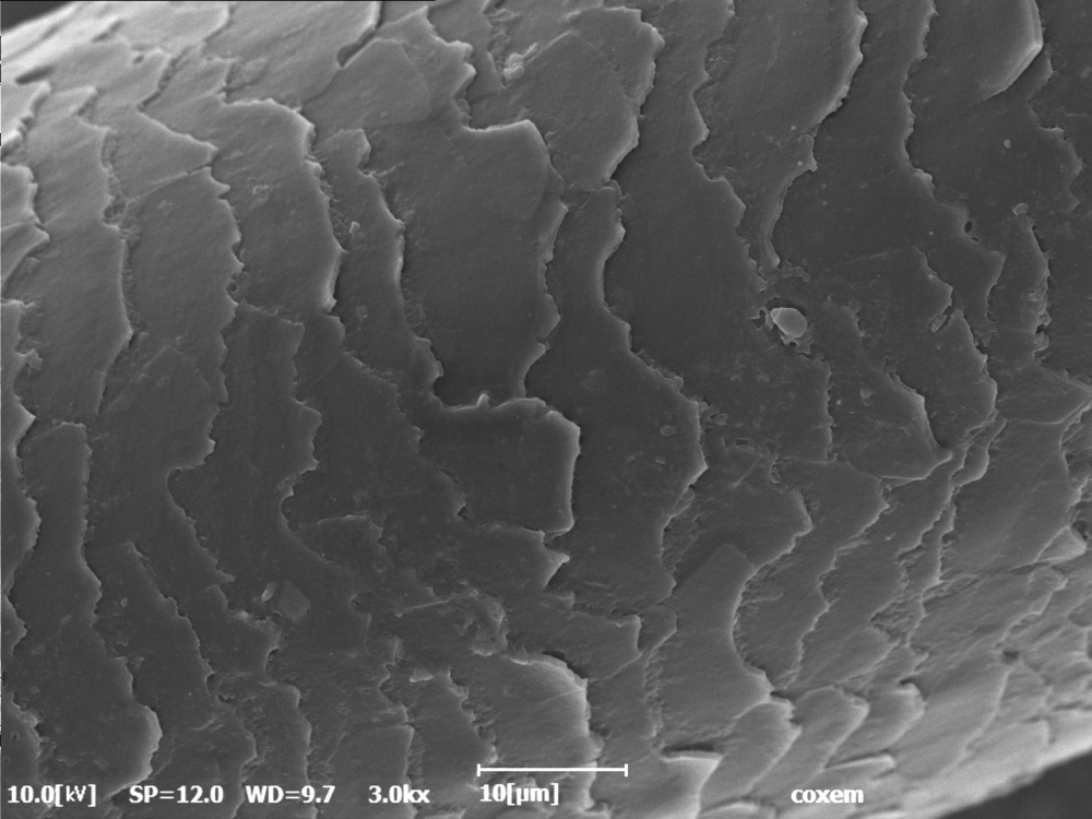
[Hair 2]
The surface of the hair was observed using SEM. The overlapping cuticle layers were clearly visible, and some regions showed lifted or cracked cuticles indicating surface damage. When the cuticle layers become rough or detached, hair tends to feel coarse and lose its natural shine.
For detailed observation of fine particles and delicate surface features on the cuticle layer, lower accelerating voltage conditions are recommended to minimize surface damage and enhance surface sensitivity.

[Hair (HV)]
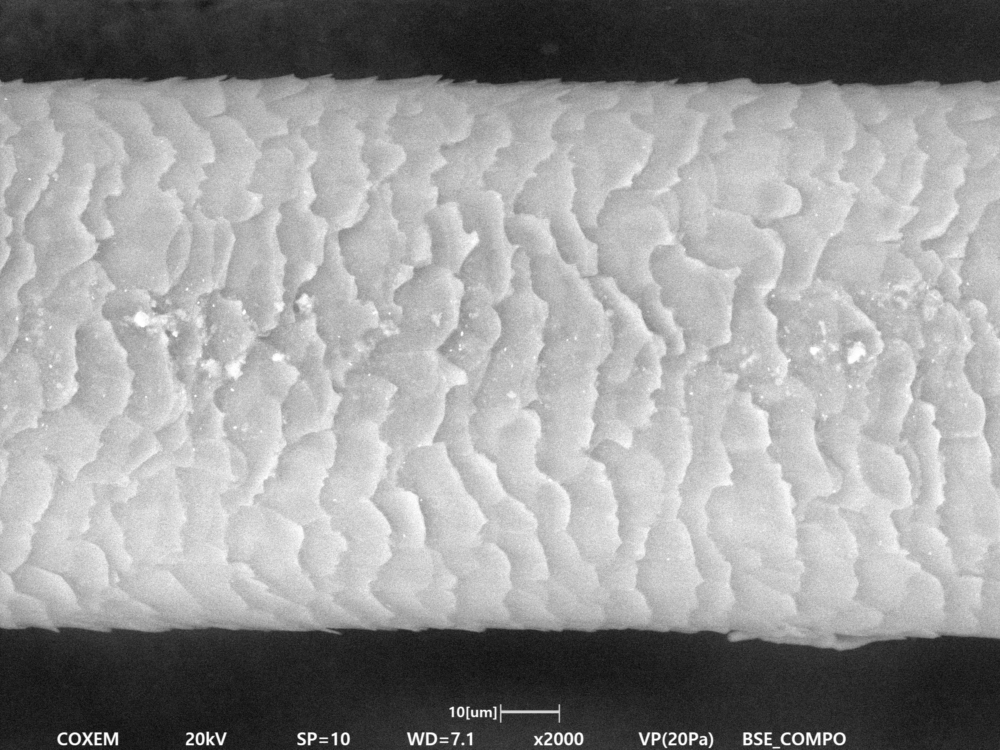
[Hair (LV)]
[Damaged Hair]

[Tip of the Hair]
[Hair Root]

 Why Large-Area SEM Analysis Matters: The Power of a Wider Field of View