Blog
Window to the Nano World



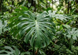
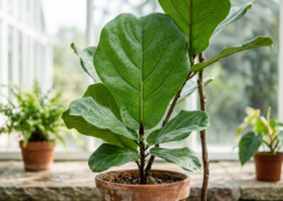
The surface of a plant leaf is far more than a simple outer layer — it is a highly specialized biological structure responsible for photosynthesis, gas exchange, moisture regulation, and protection against pathogens. The epidermis forms the outermost protective cell layer, while microscopic stomata enable carbon dioxide absorption and oxygen release.
Many plant surfaces also contain micro- and nanoscale structures that control water behavior. For example, the lotus leaf features fine surface protrusions that cause water droplets to maintain a spherical shape and roll off easily, creating a self-cleaning effect. By analyzing these natural surface structures, researchers can replicate their functions in engineered materials and industrial applications. Lotus-inspired microstructures, for instance, are widely used in superhydrophobic architectural coatings and anti-contamination treatments for solar panels.
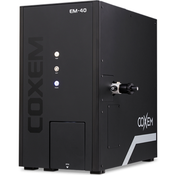
To observe these complex biological microstructures in detail, SEM analysis is essential. COXEM’s EM-40 tabletop SEM provides a fast and convenient solution for plant research, enabling detailed observation of fine cellular structures, tissues, leaf surfaces, and root cells that are difficult to resolve using conventional optical microscopy.
Living plant tissues may contain significant moisture, which can interfere with SEM vacuum conditions and reduce image quality. If a moisture-rich sample is introduced directly into the SEM chamber, unstable vacuum conditions may occur, affecting imaging performance. To prevent this, samples should be thoroughly dried before observation. Alternatively, a cooling stage can be used to freeze residual moisture, enabling stable observation in a solid state.
Because plant samples are non-conductive, conductive coating is also recommended to prevent charging during SEM imaging.
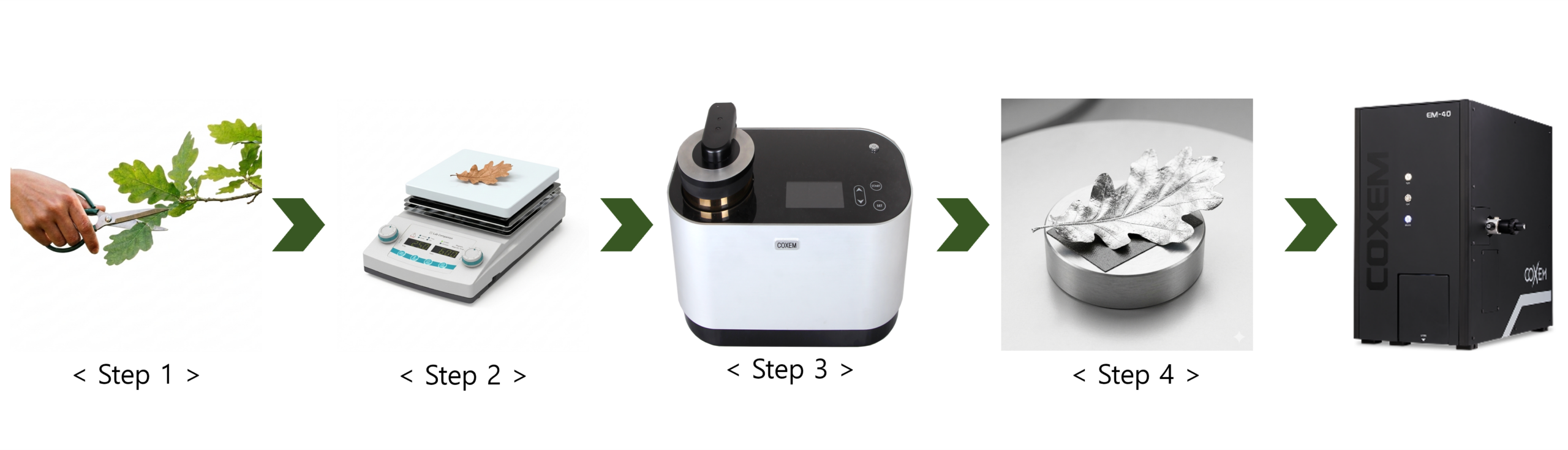
Step 1. Cut the plant sample to fit the size of the SEM holder.
Step 2. If the sample contains excess moisture, dry it at room temperature or on a hot plate.
Step 3. Mount the dried sample onto the holder using conductive materials such as carbon tape or silver paste.
Step 4. Place the mounted sample into the SPT-20 and apply Pt or Au coating.
Step 5. Load the coated sample into the EM-40 and perform SEM observation.

[Surface of a Living Leaf]
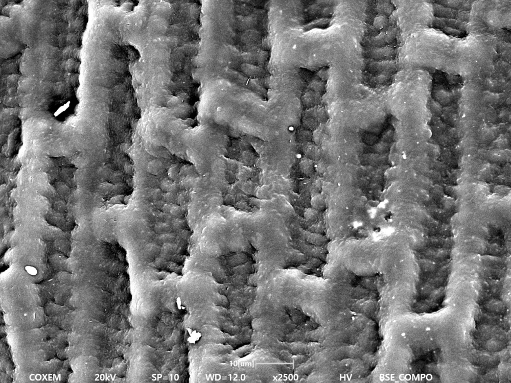
[Surface of a Dried Leaf]
The surfaces of both living and dried leaves were observed using SEM. In the living leaf, residual moisture maintained cellular turgor pressure, resulting in a firm and well-defined surface structure. In contrast, the dried leaf showed shrinkage and deformation of epidermal cells due to moisture loss, along with collapsed regions of the cuticle layer.

[Stomata of Living Leaf]
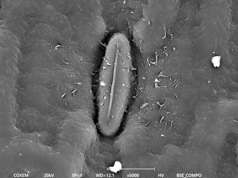
[Stomata of Dried Leaf]
Stomata on the surfaces of living and dried leaves were observed using SEM. In the living leaf, the guard cells were partially open, allowing gas exchange such as carbon dioxide intake and oxygen release through the stomatal opening.
These guard cells expand when hydrated, causing the stomata to open, and contract as moisture decreases, leading to closure. In the dried leaf, loss of moisture caused the guard cells to shrink, resulting in closed stomata.
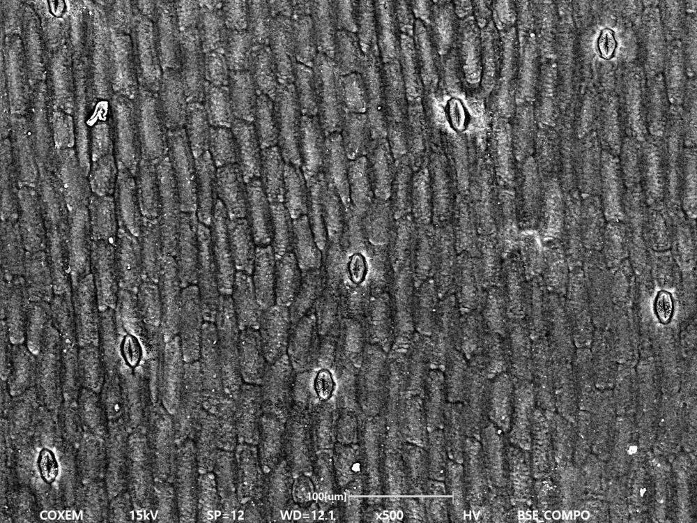
[Surface of a Leaf 1]
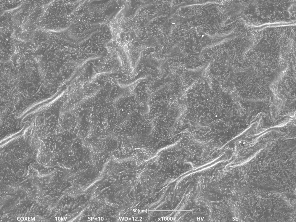
[Surface of a Leaf 2]
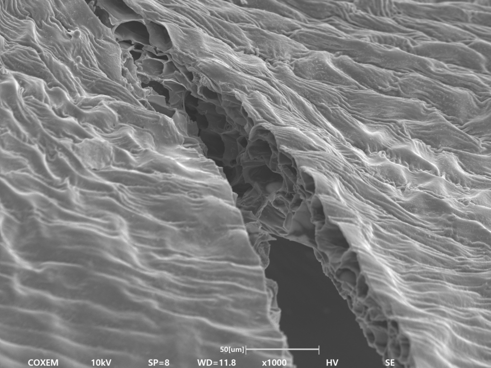
[Surface of a Leaf 3]
#Leaf #EM-40 #LifeScience #Bio

 Visualizing Microneedles: SEM Analysis of a Rising Cosmetic Ingredient